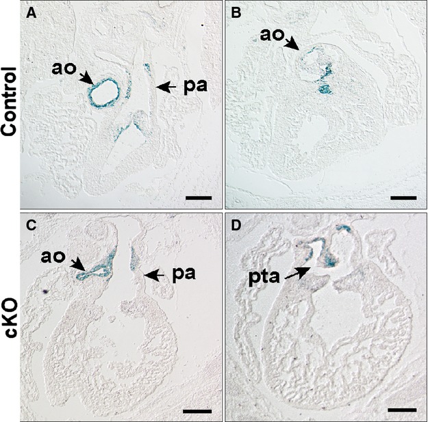
Figure 6.

NCC fate map in an E15.5 embryo with Wnt1Cre mediated inactivation of Hif‐1α. NCC fate is mapped with histological detection of LacZ activity from the Rosa26 locus. Matched sections are from anterior to posterior. In control (A and B; Hif‐1αf/f; Wnt1Cre−) and cKO (C and D; Hif‐1αf/f; Wnt1Cre+) embryos, LacZ+ cells are present in the aorta, pulmonary artery and the semilunar valves. In the control embryo LacZ+ cells are evident in the rostral portion of the ventricular septum below the aortic valve (B). The outlet septum is absent in the cKO accounting for the outlet VSD. D, LacZ+ cells are observed in cKO in the abnormally positioned aortico‐pulmonic septum (arrow), which is incomplete resulting in PTA. Scale bars: 500 μm. Ao indicates aorta; cKO, conditional knock‐out; Hif, hypoxia‐inducible transcription factor; NCC, neural crest cells; pa, pulmonary artery; PTA, persistent truncus arteriosus; VSD, ventricular septal defect.
