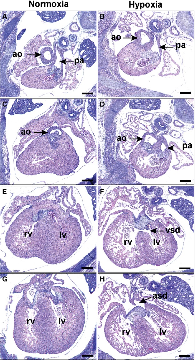
Figure 8.

Effect of maternal O2 deprivation on cardiac outlet morphogenesis. Pregnant mice were housed at 0.5 ATM (half of the normal O2 content) from E10.5 to 13.5 and returned to normal conditions until E15.5. Sections shown are from E15.5 normoxic (A, C, E, and G) and hypoxic (B, D, F, and H) embryos from anterior to posterior with respect to the heart. B and D, The aorta of the hypoxic embryo is mal‐positioned originating partly from the RVOT in DORV morphology. More posteriorly is (F) VSD and (H) ASD with persistence of the AV cushion mesenchyme. Scale bars: 500 μm. Ao indicates aorta; ASD, atrial septal defect; ATM, atmospheres; DORV, double outlet right ventricle; LV, left ventricle; pa, pulmonary artery; RV, right ventricle; VSD, ventricular septal defect.
