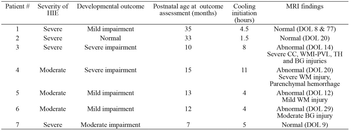
Table 2. Developmental outcome of the studied neonates in relation to the severity of HIE, time of cooling initiation from birth and MRI findings before hospital discharge. Timing of MRI is also shown.

BG: basal ganglia, CC: cerebral cortex, DOL: day of life, HIE: hypoxic-ischemic encephalopathy, MRI: magnetic resonance imaging, WM: white matter, PVL: periventricular leukomalacia, TH: thalamus.
