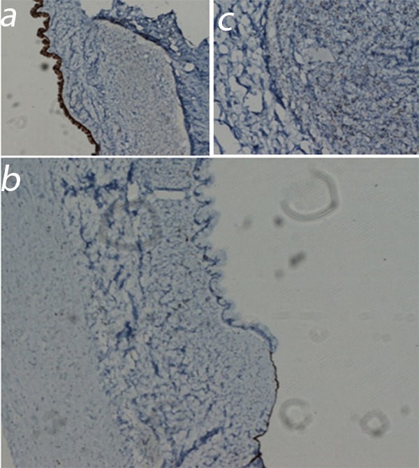
Figure 2. Immunohistochemical examination of the cysts: a) columnar mucinous cells showing positivity for CK7 (CK7, x 200), b) cuboidal cells’ positivity for calretinin (calretinin, x200), c) stromal cells’ positivity for estrogen receptor (ER, x400).

