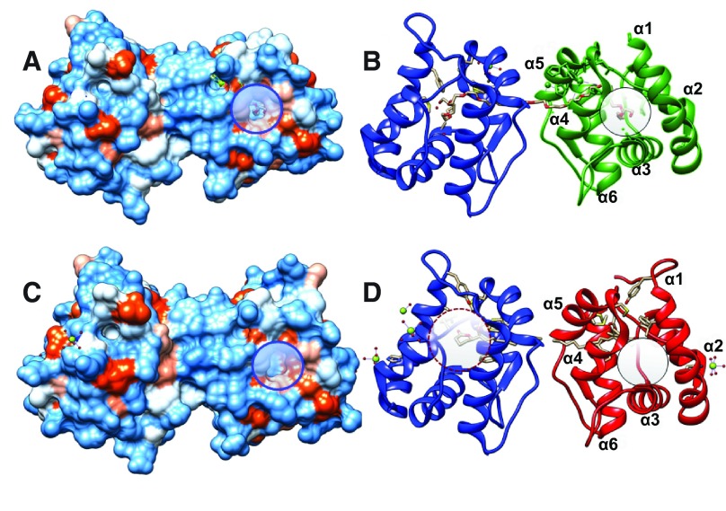
Figure 7. Structures of AaegOBP1 and CquiOBP1 bound to PEG and MOP, respectively.
( A and C) Hydrophobicity surfaces of AaegOBP1 and CquiOBP1. ( B and D) Ribbon displays of the same structures. A potential secondary binding site for MOP is highlighted with circles. It is occupied by PEG in AaegOBP1 but “empty” in CquiOBP1. The central cavity is highlighted in ( D) with a dashed circle and shows that only the polar head (lactone moiety) of MOP is housed in the core of the protein. Figure prepared with UCSF Chimera software.

