
Figure 3.
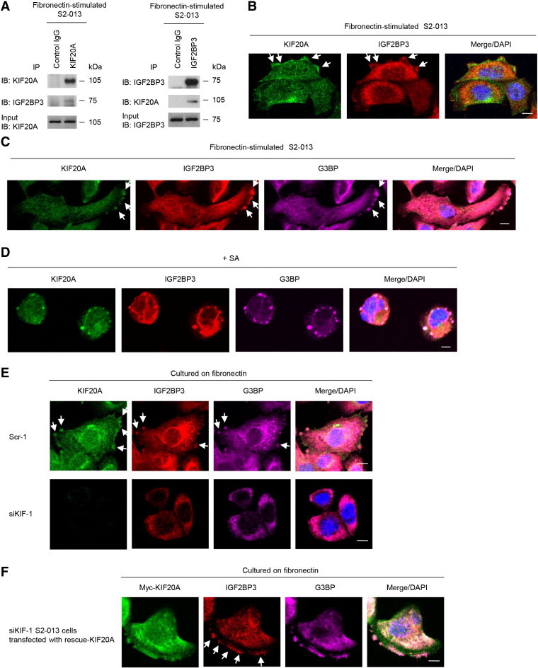
KIF20A colocalizes with IGF2BP3 in cytoplasmic RNA granules assembled in cell protrusions.
(A) Immunoprecipitation of KIF20A or IGF2BP3 from fibronectin-stimulated S2-013 cells. Immunoprecipitants were examined by Western blotting using anti-KIF20A and anti-IGF2BP3 antibodies. Mouse IgG isotype control antibody was used as an isotype control.
(B) Immunocytochemical staining of S2-013 cells cultured on fibronectin; anti-KIF20A (green) and anti-IGF2BP3 (red) antibodies were used to label endogenous proteins. Arrows, KIF20A colocalized with IGF2BP3 in cell protrusions. Blue, DAPI staining. Bar, 10 μm.
(C) S2-013 cells were incubated on fibronectin and immunocytochemically stained using anti-KIF20A (green), anti-IGF2BP3 (red), and anti-G3BP (violet) antibodies. Arrows, KIF20A colocalized with IGF2BP3 and G3BP in cell protrusions. Bar, 10 μm.
(D) S2-013 cells were exposed to 500 μM SA for 30 minutes. Immunocytochemical staining with anti-KIF20A (green), anti-IGF2BP3 (red), and anti-G3BP (violet) antibodies is shown. Blue, DAPI staining. Bar, 10 μm.
(E) Scrambled control (Scr-1) S2-013 cells or KIF20A-knockdown (siKIF-1) S2-013 cells were incubated on fibronectin and immunocytochemically stained with anti-KIF20A (green), anti-IGF2BP3 (red), and anti-G3BP (violet) antibodies. Arrows, KIF20A colocalized with IGF2BP3 and G3BP in cell protrusions. Blue, DAPI staining. Bars, 10 μm.
(F) The myc-tagged KIF20A-rescue construct was transfected into KIF20A-knockdown (siKIF-1) S2-013 cells. Forty-eight hours later, the cells were incubated on fibronectin. The cells were immunocytochemically stained with antibodies against myc (green), IGF2BP3 (red), and G3BP (violet). Arrows, reexpressed IGF2BP3 colocalized with G3BP in cell protrusions. Blue, DAPI staining. Bar, 10 μm.