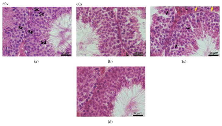
Figure 4.
Morphological analysis of mouse testis after different postfixative manipulations. Sections of adult mouse testis were stained with HE (all at 60x; magnification bar is 40 μm). (a) Control; (b) slow rehydration-dehydration stepwise replacement of ETOH-PBS-ETOH; (c) slow Re-ETOH only stepwise replacement; (d) rapid ETOH-PBS-ETOH transition. Sertoli cell (Se), spermatogonia (Sp), spermatocytes (Sc), spermatids (Sd), and Leydig cell (L). Black arrows—abnormal open spaces in seminiferous epithelium; yellow arrows—abnormal wavy and thinner basement membrane.

