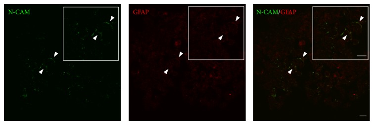
Figure 8.

Localisation of glial and neuronal cell markers in the GS-aggregates. Representative confocal images of GS-aggregates cultured under the modelled microgravity, for 2 weeks, and immunostained with anti-N-CAM and anti-GFAP antibodies (as indicated). Insets show image magnification. Scale bars, 20 μm.
