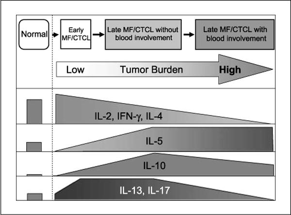
Fig. 4.

Schematic model for stages of MF/CTCL and summary of PBMC cytokine expression in normal, early MF/CTCL, and late MF/CTCL patients. Based on our cytokine expression results, functional staging of MF begins at early MF/CTCL, progresses to late MF/CTCL−B, and ends with late MF/CTCL +B. IL-2, IL-4, and IFN-γ exhibit a downward trend from early MF/CTCL to late MF/CTCL+B. IL-5 is elevated in late MF/CTCL, whereas IL-10 levels peak at late MF/CTCL−B and then decrease in late MF/CTCL+B. IL-13 and IL-17 levels increase in early MF/CTCL and late MF/CTCL−B but decrease in late MF/CTCL+B.
