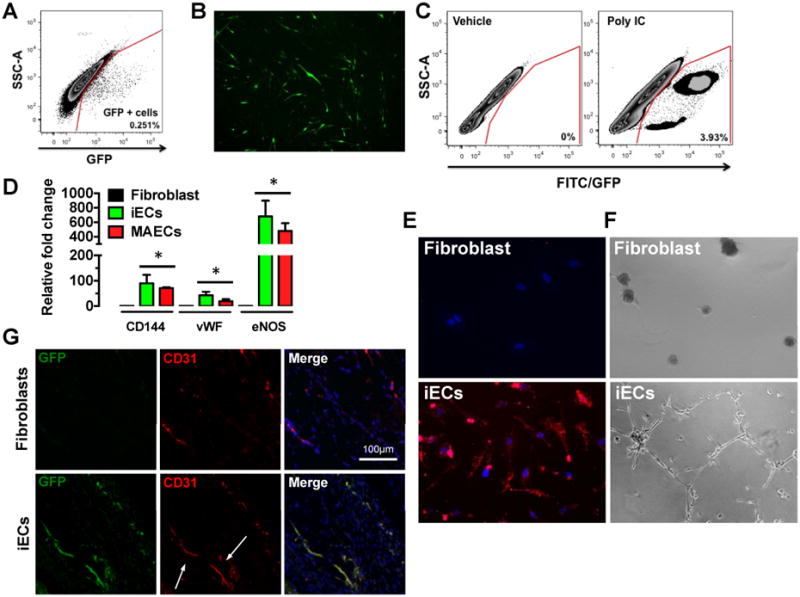
Figure 2.

Direct reprogramming of mouse tail-tip fibroblasts to functional iECs. (A) FACs plot obtained by sorting GFP positive and GFP negative cells from tail-tip (TT) fibroblasts. GFP positive cells were eliminated and GFP negative cells were collected by FACs. (B) Representative image of tail-tip fibroblasts expressing GFP at day 19 of the transdifferentiation protocol. (C) FACs plot obtained by sorting GFP positive cells at day 28 following the transdifferentiation protocol to quantify iECs. (left panel) - vehicle control; (right panel) - Poly I:C. (D) Real-Time RT-PCR for endothelial markers CD144, vWF and eNOS in miECs mouse and aortic endothelial cells (MAECs), which are comparable (n=3, done in duplicates) *p<0.05, 1-way ANOVA corrected with Bonferroni method. (E) miECs incorporate acetylated LDL compared to tail-tip fibroblasts. (F) miECs form capillary-like network compared to TT fibroblasts when seeded on matrigel. (G) miECs form blood capillaries in subcutaneous matrigel plugs as shown by CD31 staining.
