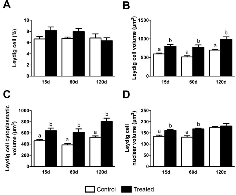
Figure 8.

Morphological alterations of mouse Leydig cells at 15, 60 and 120 days after silver nanoparticles treatment. Although the Leydig cell volume density (A) and their number per testis (B) were similar among control and treated groups, the nuclear (C) and the cytoplasmatic (D) volume of Leydig cells were significantly higher (P<0.05) in mice treated with silver nanoparticles. As a consequence, the Leydig cell volume/size (E) significantly increased (P<0.05) in treated mice. Data are presented as means ± standard error of mean (SEM). Statistical analysis was performed using one-way ANOVA. Letters denote post-hoc test differences.
