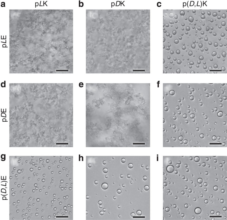
Figure 1. Optical micrographs of polyelectrolyte complexes.
Bright-field optical micrographs showing the liquid coacervates or solid precipitates resulting from the stoichiometric electrostatic complexation of L, D, or racemic (D,L) poly(lysine) with L, D or racemic (D,L) poly(glutamic acid) at a total residue concentration of 6 and 100 mM NaCl. Complexes are formed from (a) pLK+pLE, (b) pDK+pLE, (c) p(D,L)K+pLE, (d) pLK+pDE, (e) pDK+pDE, (f) p(D,L)K+pDE, (g) pLK+p(D,L)E, (h) pDK+p(D,L)E, (i) p(D,L)K+p(D,L)E. Liquid coacervate droplets are only observed during complexation involving a racemic polymer. Scale bars, 25 μm.

