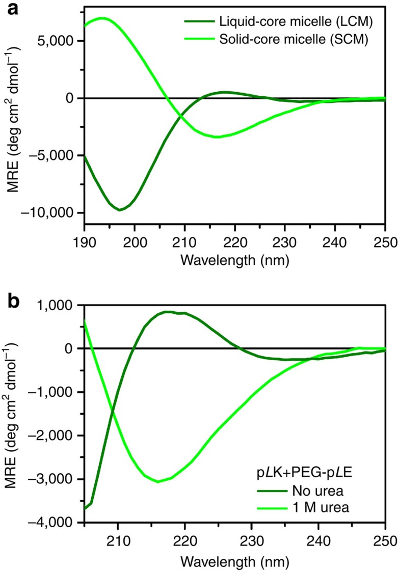
Figure 4. Secondary structure of micellar complexes.
CD spectra for (a) LCM (dark green) and SCM (light green) micelles formed from PEG-pLK+p(D,L)E and PEG-pLK+pLE, respectively, and (b) SCMs in the absence (light green) and presence of 1 M urea (dark green), respectively. LCMs show a random coil structure, while SCMs display β-sheet character in the absence of urea, but convert to a random coil structure in the presence of 1 M urea, suggestive of a LCM. LCMs were prepared at a polymer concentration of 0.01 mM total polymer concentration, while SCMs were prepared at 0.0125, mM with an average N=100 for the micelles in a, and both LCMs and SCMs were prepared at a total polymer concentration of 0.04 mM with an average N=50 for b.

