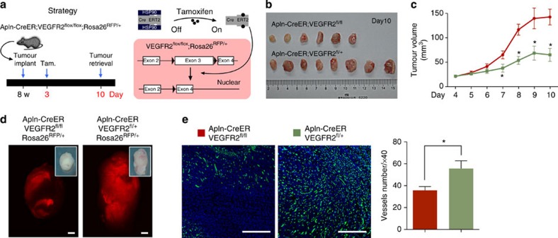
Figure 7. Apln-CreER-mediated genetic ablation of VEGFR2.
(a) Schematic showing the strategy and principle of induced VEGFR2 ablation in Apln+ cells. Tam., tamoxifen treatment. (b) Picture of tumours from Apln-CreER;VEGFR2fl/fl and littermate controls Apln-CreER;VEGFR2fl/+ mice. (c) Quantification of tumour volume (length × width2/2 (mm3)) at different times following implantation. Student’s t-test was used to analyse differences and values are shown as means±s.e.m.; *P<0.05; n=6–8 for each time point. (d) Whole mount view of RFP+ tumours. (e) Vessel detection by PECAM staining and quantification of vessel number in different groups. Student’s t-test was used to analyse differences and values are shown as means±s.e.m.; *P<0.05; n=6. Scale bars, 1 mm (d); 0.5 mm (e).

