
Figure 6.
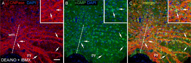
NO stimulates cGMP accumulation in oligodendrocytes in slices of 14-day-old rat brainstem. The slices were exposed to DEA/NO (100 μM, 5 min) in the presence of IBMX (1 mM). Images are from coronal sections at the level of the XII nerve, stained for (A) CNPase (red), (B) cGMP (green) and DAPI (blue). (C) overlay of A and B. Key: broken line, the mid line; py, pyramid; wm, white matter; arrows, oligodendrocytes. Insets are enlargements centred on the two arrowed cells near the midline. Scale (A) = 50 μm or 25 μm (insets). [Color figure can be viewed in the online issue, which is available at http://wileyonlinelibrary.com.]