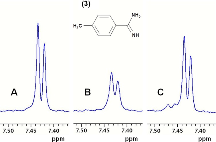
Fig 3. Ligand detected one dimensional NMR spectroscopy.
Part of 1H T1rho NMR spectra of p-methyl-benzamidine (pMeBza): (A) 100 μM pMeBza in buffer; (B) after addition of 5 μM FXIa CD; and (C) after addition of 10 μM compound 2. Shown is the doublet from the protons in the ortho position. The extra signals in C are from the inhibitor.

