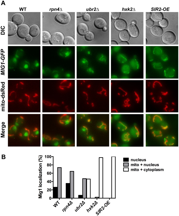
Figure 8. Mig1 co-localizes with the mitochondria in cells with increased proteasome abundance, in the absence of HXK2 and upon overexpression of SIR2.
(A) The strains indicated carrying Mig1 with a C-terminally fused GFP tag were co-transfected with the plasmid mito-dsRED, carrying a red fluorescing mitochondrial marker. Co-localization of Mig1 with the mitochondria is visualized via merging of the Mig1 and the mitochondrial signals in living cells during early logarithmic growth. Projected sequential Z-stacks fluorescence images are presented. DIC: differential interference contrast. Mig1 localization of ~300 cells for each strain was analyzed and presented in (B).

