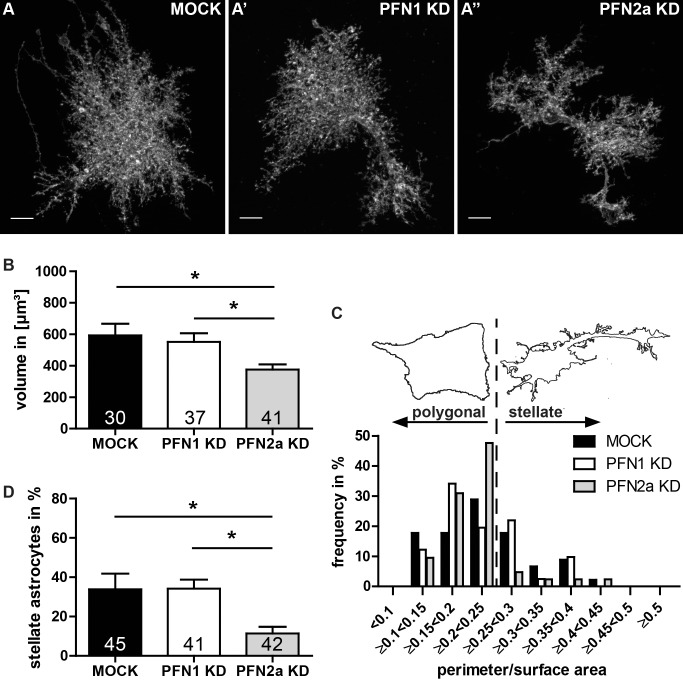
Fig 3. PFN2a modulates morphology of astrocytes in organotypic slices and dissociated cultures.
(A-A”) Morphology of astrocytes in cortical organotypic slice cultures is similar to the in vivo appearance. Images of representative astrocytes (A) MOCK transfected, expressing a PFN1 specific shRNA (A’) and a PFN2a specific shRNA (A”), respectively. Scale bar: 10 μm. (B) Statistical analysis of the volume of astrocytes from cortical slice cultures expressing the shRNA specific for PFN2a (375.6±33.2 μm²) displayed a significant reduction of their volume compared to control (592.6±74.3 μm²) or PFN1 knockdown (551.8±54.4 μm²) astrocytes. (C) Categorization of astrocytes from dissociated cultures into 2 groups: stellate and polygonal. Astrocytes were subdivided into different classes according to their ratio between cell perimeter and cell area. Subsequently, cells with a ratio above 0.25 were added up to achieve the percentage of stellate astrocytes, the remaining were totaled to the group of polygonal astrocytes. The border between these two groups is indicated as dashed line. (D) The amount of stellate cells in dissociated cultures is significantly reduced in the group of PFN2a reduced cells. Quantitative data obtained from 3 independent experiments was tested for significance by one-way ANOVA followed by a post-hoc Tukey’s Multiple Comparison Test. Significance is indicated as follows *p<0.05; **p<0.01; ***p<0.001. Data are shown as mean ± SEM.

