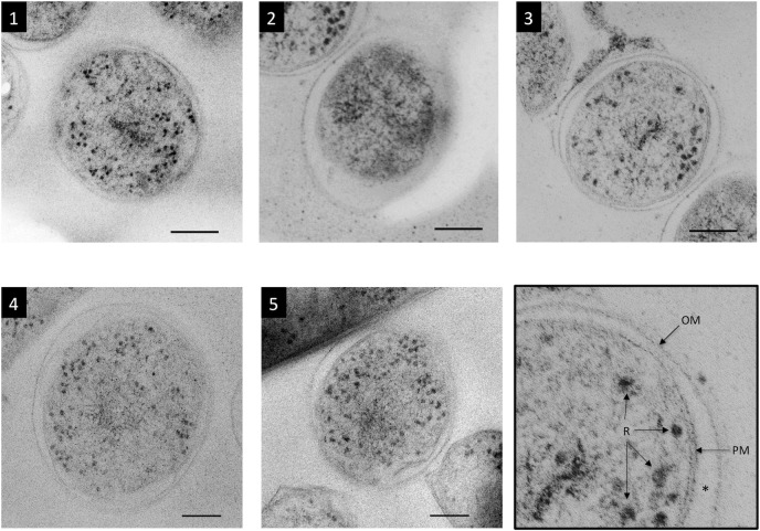
Figure 1. TEM images of five cross-sectioned M. tuberculosis cells.
Cells were cut in the middle. The outer membrane (OM), periplasm (asterisk), plasma membrane (PM), and ribosomes (R) are visible as shown in the bottom right panel (enlarged image of cell 3). The cytoplasm of cell 2 appeared to have degraded, as evidenced by its dark color and fewer ribosomes. Cell 3 can be seen to the upper left of cell 2. Bar: 100 nm.

