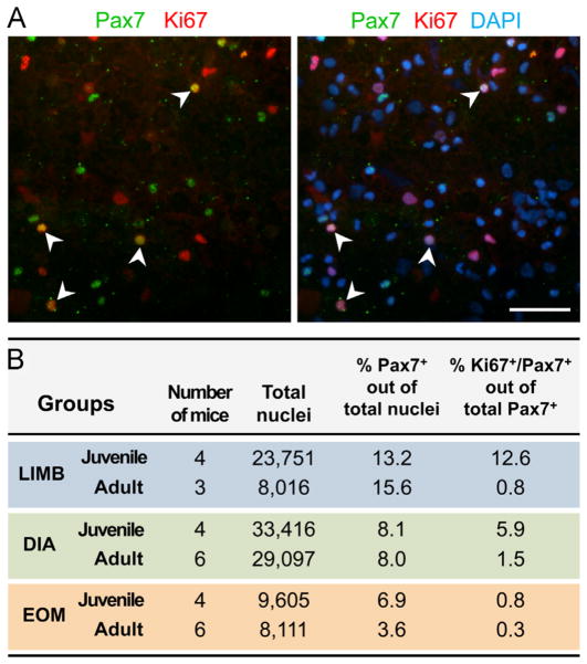
Fig. 4.
Assessment of in-vivo proliferative activity of SCs. Unsorted cell preparations obtained after Pronase digestion of LIMB, DIA and EOM of adult (4–6 month old) and juvenile (3-week old) mice were subjected to cytocentrifugation, followed by Pax7/Ki67 double immunostaining combined with DAPI counter-staining. (A) Image shown is of a preparation from LIMB of juvenile mice, which contains the highest number of Ki67+/Pax7+ double-labeled cells. (B) SCs were identified based on Pax7 immunostaining and their proliferative activity was determined by calculating the percent of cells double-immunolabelled for Pax7 and Ki67 (Ki67+/Pax7+ cells) out of all Pax7+ cells. Preparations of spleen cells were used as a highly proliferative control for Ki67 immunolabeling (Ki67+ cells: 48.1% out of 7998 and 18.0% out of 4324 total nuclei, in juvenile and adult mice, respectively); these cells were also found negative for Pax7. Scale bar, 50 μm.

