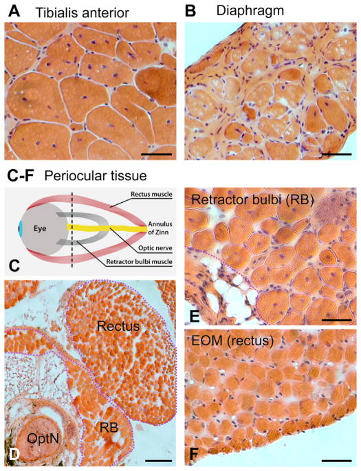
Fig. 5.
H&E stained cross sections of tissue preparations from mdx4cv mice. (A) TA, (B) DIA, and (C–F) periocular tissues from 1 year old mice. (C) Schematic of a periocular tissue preparation that comprises the EOMs, the retractor bulbi (RB) and the optic nerve (OptN), harvested together with the eyeball. The black dotted line indicates the level of the sections shown in: (D) periocular tissue (low magnification), (E) RB and (F) EOM (high magnification). Scale bars, 50 μm (A and B) and (E and F), 200 μm (D).

