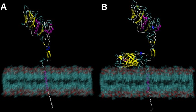
FIG 4.

Crystal structure model of HE and HE-GFP chimeric proteins. (A) Cartoon representation of crystal structure model of the ISAV HE protein obtained by homology modeling using the HEF protein from influenza C virus (PDB entry 1FLC). (B) Modeling of the HE protein structure of ISAV901_09 containing EGFP inserted within HPR. Protein structures are modeled embedded in a lipid membrane. The secondary structures represent the β-sheet (yellow), alpha helix (purple), short alpha helix (blue), unstructured chain (gray), and loop chain (green/cyan).
