
Figure 3.
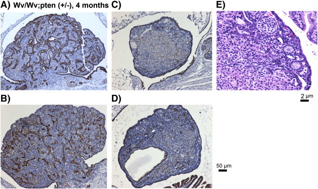
Ovarian morphological features of Wv/Wv; Pten (+/−) mutant mice. Representative CK8 staining (of epithelial cells) of four examples of ovaries from four 4-month-old Wv/Wv; Pten (+/−) mice is shown. Two ovaries (A, B) are similar to those of Wv/Wv mice, and the other two ovaries shown (C, D) exhibit a reduced tumor lesion. An example of H&E staining of the ovary with reduced tumor lesion is shown at higher magnification to visualize the presence of follicle structures (E).