
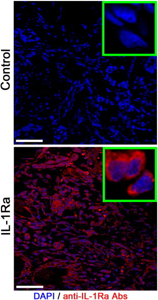
Figure 1.
Confocal microscopy of the C6 glioma following i.v. administration of the IL-1Ra. Nuclei were stained with DAPI (blue). IL-1Ra was detected with monoclonal antibodies conjugated with Alexa Fluor 555 (red). Scale bar, 75 μm.
Official websites use .gov
A
.gov website belongs to an official
government organization in the United States.
Secure .gov websites use HTTPS
A lock (
) or https:// means you've safely
connected to the .gov website. Share sensitive
information only on official, secure websites.
Confocal microscopy of the C6 glioma following i.v. administration of the IL-1Ra. Nuclei were stained with DAPI (blue). IL-1Ra was detected with monoclonal antibodies conjugated with Alexa Fluor 555 (red). Scale bar, 75 μm.