Figure 2.
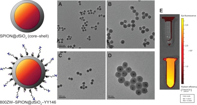
Schematic and TEM/NIRF pictures of actual structures before and after surface modification.
Notes: (A) A TEM image of SPION@dSiO2 (core–shell) before surface modification (scale bar =100 nm). (B) A zoomed-in TEM image of SPION@dSiO2 (core–shell) before surface modification (scale bar =50 nm). (C) A TEM image of 800ZW–SPION@dSiO2–YY146 (scale bar =100 nm). (D) A magnified TEM image of 800ZW–SPION@dSiO2–YY146 (scale bar =50 nm). (E) An NIRF imaging of nanoparticle (upper) of SPION@dSiO2 and (down) 800ZW–SPION@dSiO2–YY146 in PBS solution.
Abbreviations: TEM, transmission electron microscopy; SPION, superparamagnetic iron oxide nanoparticles; NIRF, near infra-red fluorescence; PBS, phosphate-buffered saline; Min, minimum; Max, maximum.

