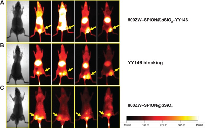
Figure 6.
NIRF imaging.
Notes: Serial coronal NIRF images of MKN45 tumor-bearing mice at different time points postinjection of (A) 800ZW–SPION@dSiO2–YY146, (B) 800ZW–SPI N@dSiO2 with a blocking dose of YY146, or (C) 800ZW–SPION@dSiO2. Tumors are indicated by yellow arrowheads.
Abbreviations: NIRF, near infra-red fluorescence; SPION, superparamagnetic iron oxide nanoparticles.

