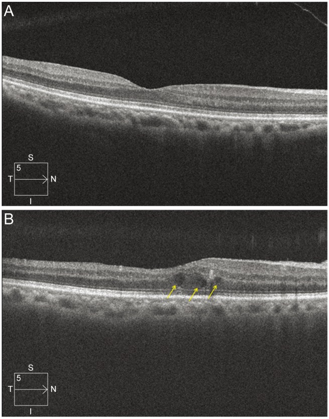
Fig. 2.

Macular scan image of the same patient in Fig. 1. (A) Preoperative macular cube scan image. The central subfield retinal thickness (CRT) was 267 µm, and the fovea did not show retinal thickening or cystoid change. (B) Image of macular cube scan 1 month after surgery. CRT increased to 324 µm. The normal foveal depression disappeared, and the images show cystoid change at the fovea (arrows). I = inferior; N = nasal; S = superior; T = temporal.
