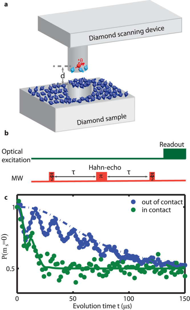
Figure 1. Detecting surface electron spin ensembles using a scanning magnetometer composed of a single nitrogen-vacancy (NV) spin.

(a): Schematic of the experiment. The NV spin resides in a nanopillar fabricated on a diamond scanning platform, with distance d from the sample surface. The angle between the normal direction of the surface and the quantization axis of the NV spin θ is imposed by crystallographic directions. The NV spin is optically initialized and read out from above. A nearby antenna is used to apply microwave pulses to manipulate the NV spin state coherently. The diamond sample contains a mesa that is 200 nm in diameter and 50 nm in height, with paramagnetic impurities on the surface. The NV spin is decohered by both the surface spins on the nanopillar and on the sample. (b): Hahn-echo experimental protocol (with evolution time t = 2τ). (c): Measurements of the NV spin state (ms = 0) in the scanning nanopillar after application of the experimental protocol in b, showing strong reduction of NV spin coherence when the nanopillar is in contact with the sample compared with it retracted 4 μm from the surface of the sample27. The blue solid line shows the stretched exponential fit with Gaussian shaped peaks for the collapses and revivals from nearby 13C nuclear spins. The blue dotted dash line and the green solid line show the fits to the spin bath model as described in the text.
