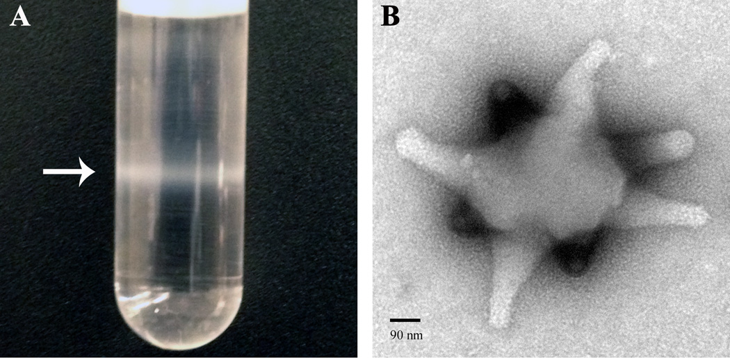
Figure 1. Virulence factors from Leptopilina species.
(A) An opaque band of concentrated purified VLPs (arrow) is visualized after ultracentrifugation of venom gland extract in a nycodenz gradient. (B) A uranyl acetate stained VLP, visualized by the negative stain transmission electron microscope (Zeiss 902) method. Downward-facing extensions from VLP body (i.e., “spikes”) are in a different focal plane and stain more intensely than upward-facing ones. VLP spikes appear to widen slightly at their termini. Spiked VLPs have been reported from L. heterotoma, L, victoriae, and L. boulardi [7,8,10]. Abundant proteins from VLPs of the L. heterotoma and L. victoriae sister species are produced in secretory cells of the wasp’s venom gland. VLP proteins and precursors transit through an extensive and conserved canal system within the venom gland, localize to specific VLP regions, and enter host hemocytes post infection [9,61]. The biological nature of VLPs (i.e., whether they are true viruses, virus-like, or simply secretions of wasp’s venom glands), their macromolecular constituents, and precise modes of action on the host’s immune system and development remain a significant challenge.

