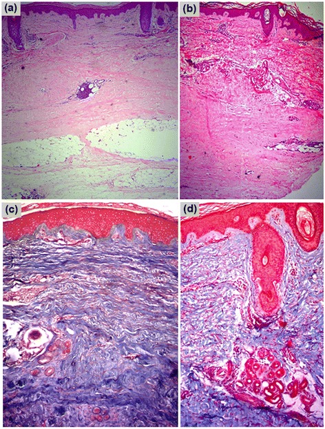
Figure 3.

Histologic changes before and after treatment. (a) Pretreatment fibrosis, well-defined pockets of perivascular inflammation in the dermis and subcutaneous cellular tissue. (b) Posttreatment biopsy, collagen loosening of compaction in the superficial and middle reticular dermis with recovery of the annexes. (c) Masson’s trichromic stain before treatment. (d) Posttreatment biopsy showing the changes described previously (original magnification, 40×).
