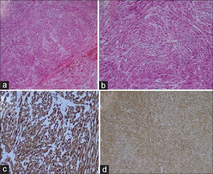
Figure 3.

(a) H and E, demonstrating, tumor composed of spindle cells (H and E, ×40), (b) H and E, demonstrating, tumor composed of spindle cells (H and E, ×100), (c) immunohistochemistry showing, desmin (×200), (d) immunohistochemistry showing, actin (×40)
