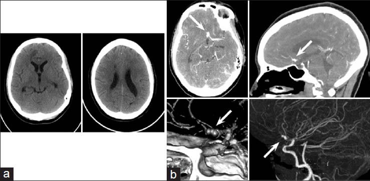
Figure 1.

Computed tomography: (a) Axial noncontrast head CT demonstrating minimal interhemispheric SAH and multiple areas of acute infarct, most prominently in the anterior cerebral artery distribution. (b) Axial CTA with sagittal and 3D reconstructions demonstrating an 8 × 5 mm bilobed saccular aneurysm located at the junction of the left anterior cerebral anterior and anterior communicating artery (arrows)
