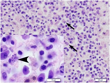
Figure 3.

Histological section of a lymph node from female D. The medulla contained numerous plasma cells and macrophages. Within the cytoplasm of some of the macrophages there were variably sized oval to round particles (arrows and arrow head). Kinetoplasts typical for Leishmania sp. were not seen. Hematoxylin and eosin (HE) stain.
