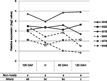
Figure 5.

Expression patterns of MdPME2 for each individual hybrid as determined by microarray analysis at four developmental stages (100 DAF, H, 60 DAH and 120 DAH). Hybrids displayed two different expression patterns: high and stable PME gene expression in non-mealy hybrids (bold lines) contrary to lowest and decreasing gene expression in mealy hybrids (dash lines). The transcript levels were normalized with the Lowess method. Normalized intensities (i.e. expression levels) were then subtracted from the background. Table refers to Kruskall-Wallis results between mealy and non-mealy hybrids at each time point.
