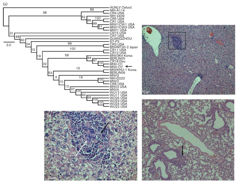
Fig. 1.
MNV phylogenetic tree including MNV-O7 and pathology caused by natural infection. (a) Full-length genomic MNV nucleotide sequences were used to construct a maximum-likelihood phylogenetic tree with PhyML and 100 bootstrap interactions. The bootstrap values are indicated on the branches. Bar, 2 % nucleotide divergence. (b–d) Representative pathology in liver (b, c) and lung (d) sections from naturally infected Stat1−/− mice from which MNV-O7 was derived. (b) A focus of inflammation in the liver parenchyma that is magnified in (c) to show the presence of inflammatory cells (macrophages shown by the white arrow) and necrotic (black arrow) cells. (d) General infiltration of the lung; the arrow indicates a localized focus of inflammation associated with a bronchiole and blood vessel. Original magnification, ×10 (b, d) and ×40 (c).

