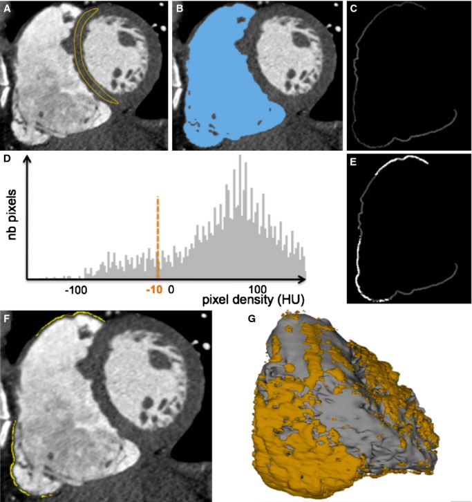
Figure 1.

Method for the segmentation of myocardial fat from MDCT images. On contrast‐enhanced ECG‐gated cardiac images reformatted in short axis, a region of interest is drawn on the interventricular septum to assess normal myocardial density (A). The RV endocardium is automatically segmented using region‐growing segmentation with a lower density cut‐off 3 SD above mean myocardial density (B). A 2‐mm‐thick RV free wall is derived from this segmentation using a dilatation operator (C). Myocardial fat is segmented on the histogram as pixels with density <−10 HU (D through F). Segmented images are then used to compute 3D objects compatible with 3D‐EAM systems (G). 3D‐EAM indicates 3‐dimensional electroanatomic mapping; MDCT, multidetector computed tomography; RV, right ventricle.
