Figure 2.

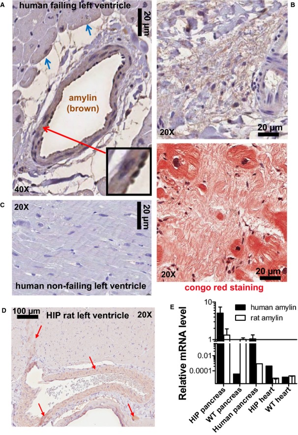
Amylin incorporates in blood vessels and parenchyma in failing hearts from diabetic humans and HIP rat hearts. A, Immunohistochemistry analysis of failing hearts from T2D patients with an antiamylin antibody shows amylin deposits (brown) incorporated in the blood vessel wall, perivascular space, and on the sarcolemma of neighboring cardiac myocytes (blue arrows). The inset shows amylin deposition in the basement membranes and co‐localization with neutrophils. B, Amylin (brown) is present in atherosclerotic lesions. Staining of the next section with Congo red (lower panel) indicates that amylin deposition may form amyloid‐like structures (pink) within myocardial interstices. C, Cardiac parenchyma from nonfailing human hearts shows no amylin deposits. D, Immunohistochemistry with an antiamylin antibody shows that amylin (brown; red arrows) incorporates into blood vessel walls and heart parenchyma in HIP rat. E, Quantitative real‐time PCR shows no presence of human amylin mRNA in the HIP rat heart. Note the logarithmic scale and scale break. N=8 HIP rat heart samples and N=4 human pancreas and rat samples were analyzed in this study. WT rat heart samples (N=8) served as negative controls. HIP indicates human amylin in the pancreas; PCR, polymerase chain reaction; T2D, type 2 diabetes; WT, wild‐type.
