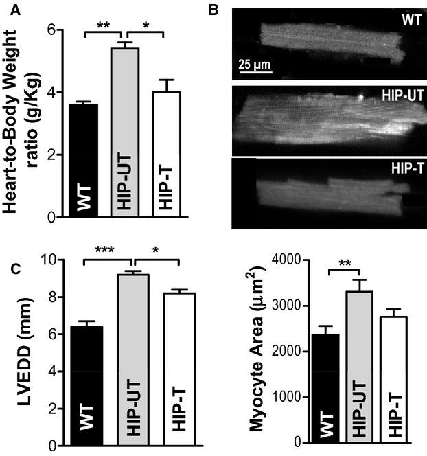
Figure 9.

Treatment reduces cardiac hypertrophy and left‐ventricular dilation in HIP rats. A, Heart‐weight‐to‐body‐weight ratio in WT, untreated HIP, and treated HIP rats. B, Top—representative examples of myocytes from WT, HIP‐UT, and HIP‐T groups; Bottom—average surface area (length×width) for WT, untreated HIP, and treated HIP rat myocytes. C, Left‐ventricular end‐diastolic diameter in WT, untreated HIP, and treated HIP rats at the end of the 13‐week study period. *P<0.05, **P<0.01, ***P<0.001. HIP indicates human amylin in the pancreas; LVEDD, left‐ventricular end‐diastolic diameter; WT, wild‐type.
