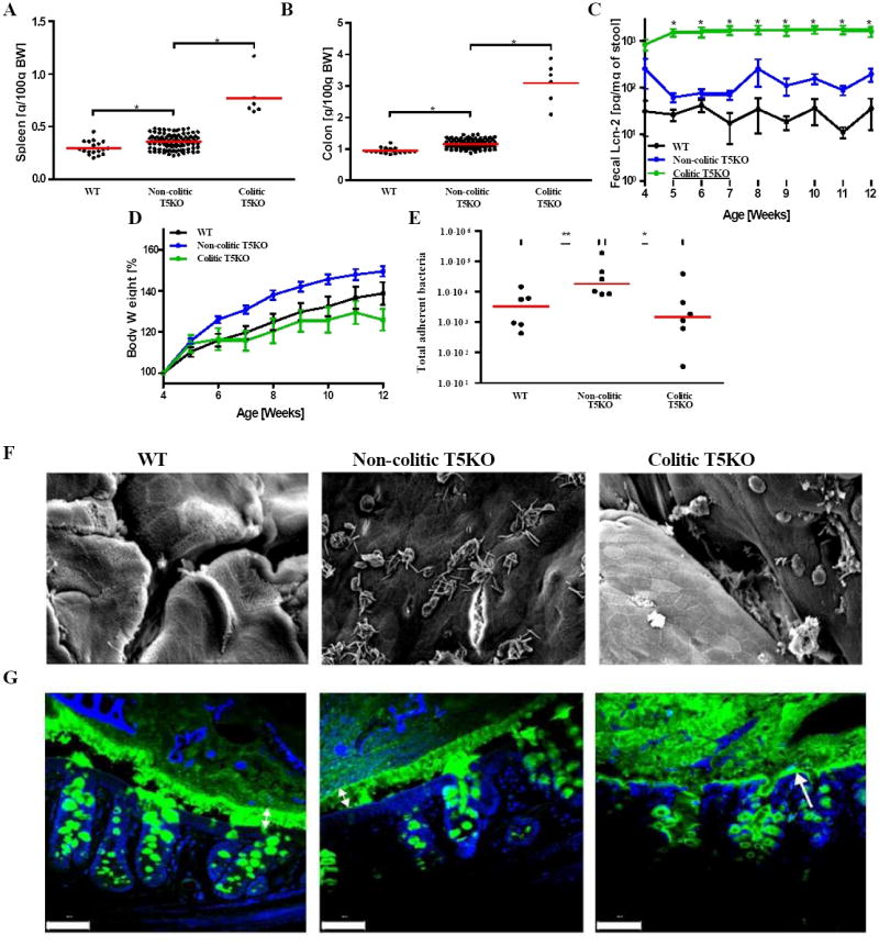
Figure 1. Development of spontaneous inflammation and microbial dysbiosis in T5KO mice.

Wild-type (n=18) and T5KO mice (n=110) were housed for 12 weeks in the animal facility to track spontaneous colitic mice (defined as described in Methods). (A) Following euthanasia, spleen was isolated and mass measured. (B) Colon mass. (C) Stool was collected weekly after weaning and diluted in 500 μL of PBS. Then, supernatant was assayed for lipocalin-2 (Lcn-2) expression by ELISA. (D) Body mass was monitored weekly from week 4 to week 12. (E) Colon was washed carefully with PBS to remove any stool and bacterial DNA was isolated. Total adherent bacteria was measured by quantitative PCR analysis using universal 16S rRNA primers. (F) Representative electron microscopy observation of colon (magnification: 1500X). (G) Muc 2 mucin immunostaining - green. Nuclei were stained using DAPI - blue (Scale bar = 20 μM). The data on panels E-G is representative of 2 independent experiments.* p<0.05.
