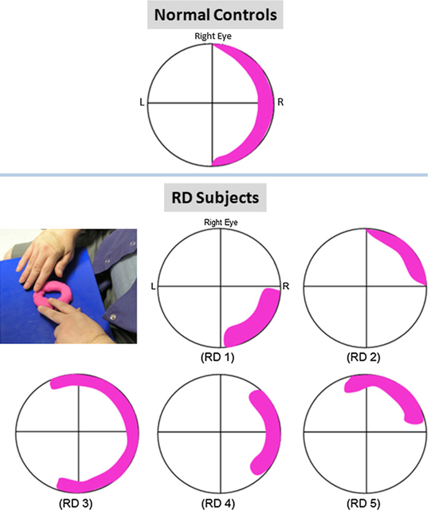
Fig. 3.
Phosphene sensation during transcorneal electrical stimulation. Pink overlay on the visual field grid indicates location and shape of phosphene reported by the subjects. (Top) cresent-shaped phosphene reported by all five normal controls; (Middle and bottom rows) phosphene sensation reported by the five retinal degenerative subjects; (Left, middle row) photograph of one retinal degenerative subject describing phosphene sensation using Play-Doh.

