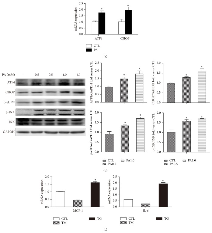
Figure 1.
ER stress is induced by palmitate and contributes to increase MCP-1 and IL-6 expression. (a) Mature 3T3-L1 adipocytes were treated with 0.5% BSA or BSA-conjugated PA (0.5 mM) for 12 h. mRNA of ATF4 and CHOP measured by qPCR. (b) Western blot of indicated ER stress markers with or without PA (0.5 or 1.0 mM) treatment. Representative blots and quantifications are shown. (c) mRNA levels of MCP-1 and IL-6 with or without the ER stressors tunicamycin (TM, 1 μg/mL) or thapsigargin (TG, 1 μM) measured by qPCR. Results are mean ± SEM of three to five separate experiments. * P < 0.05 versus nontreatment group (0.5% BSA).

