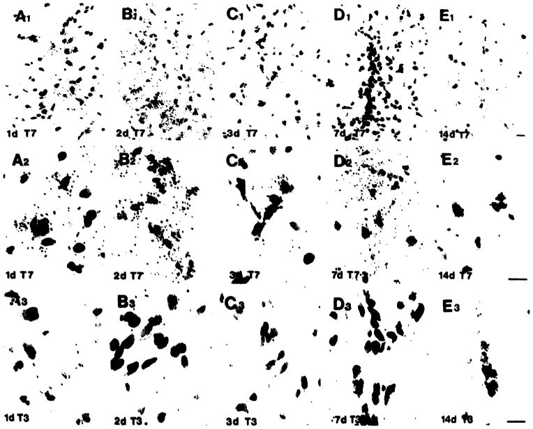
Fig. 4.
High magnification bright field micrographs of cells in the margin of the wound. TGFβ1 mRNA (T7) is observed associated with selective cells in the margin of the lesion at 1 day, (panel A) 2 days (panel B), 3 days (panel C), 7 days (panel D) and 14 days (panel E) after the lesion but not in cells hybridized with the sense strand (T3, row 3). Initial signs of the glial scar are seen at 3 days and are well defined by 7 days, at which time the mRNA appears less diffuse. High magnification reveals that these cells are typically glial cells (row 2). Bar = 10 μm.

