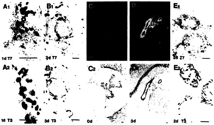
Fig. 6.
TGFβ1 mRNA in the meninges, hippocampal fissure and choroid plexus after injury. Intense TGFβ1 mRNA (T7) is observed in the meninges (panel A) and hippocampal fissure (panel B) but not in the control sections hybridized with the sense strand (T3). In the choroid plexus, TGFβ1 mRNA is almost non-detectable in uninjured animals (0 day, C1) but very intense following injury (panels D1, E1). Control sections show no signal (T3). Bright field of low-power micrographs are shown in panels C2 and D2. Bar = 25 μm.

