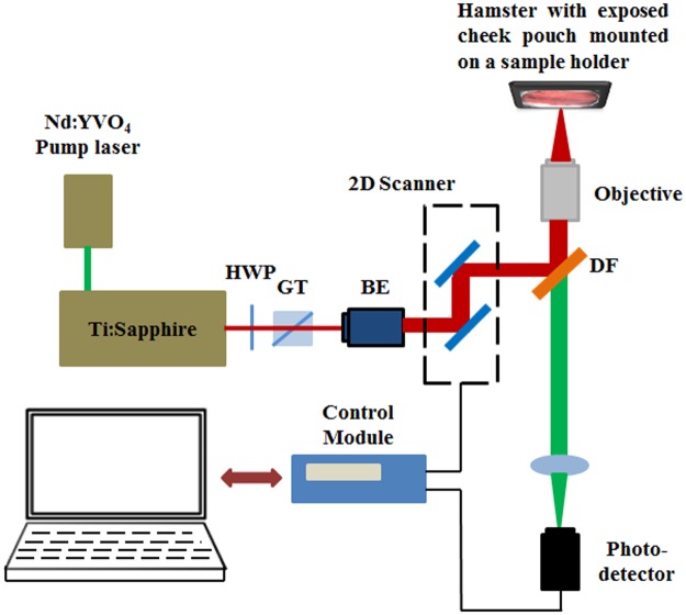
Fig 1. Schematic describing the experimental setup for in-vivo multiphoton autofluorescence microscopy (MPAM) and second harmonic generation microscopy (SHGM).
HWP: half wave plate; GT: Glans Thompson prism; BE: beam expander; DF: dichroic filter. The hamster is placed on the stage of the inverted microscope with the buccal pouch exposed and facing the objective.

