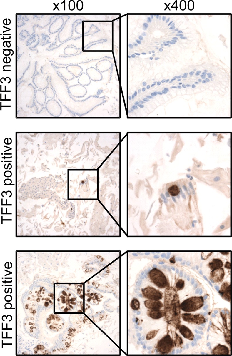
Figure 3. TFF3 immunohistochemical staining of Cytosponge samples.

TFF3 staining was performed on all Cytosponge samples to test the sensitivity and specificity of the Cytosponge-TFF3 test for diagnosing BE. TFF3 was scored in a binary fashion, with samples with one or more TFF3-positive goblet cells being classed as positive. Shown are immunohistochemical images illustrating examples of TFF3-negative and-positive staining at low magnification (100×) and high magnification (400×).
