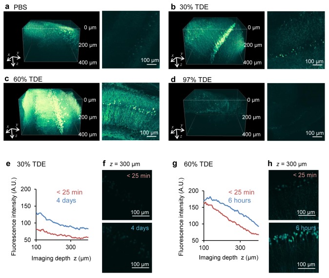
Figure 3. Enhancement of the penetration depth on two-photon microscopy.
(a–d) Images of YFP-expressing neurons in the hippocampal slices of thy1-YFP-H mouse in PBS (a) and after 2 h of immersion in 30% TDE (b), 60% TDE (c), 97% TDE (d) solution. Left, three-dimensional reconstructed images; right, xy images at a depth of 200 µm from the slice surface. (e, f) Fluorescence intensities immediately (within 25 min) and a long time (4 days) after immersion in 30% TDE solution. (e) Plot of the mean intensity of xy images against the depth from the surface; (f) Maximum projection images within 25 min and after 4 days of immersion in 30% TDE solution. (g, h) Fluorescence intensities immediately (within 25 min) and a few hours (6 h) after immersion in 60% TDE solution. (g) Plot of the mean intensity of xy images against the depth from the surface; (h) Maximum projection images within 25 min and after 6 h of immersion in 60% TDE solution.

