
Figure 5. Images of dendritic spines along a single hippocampal neuron.
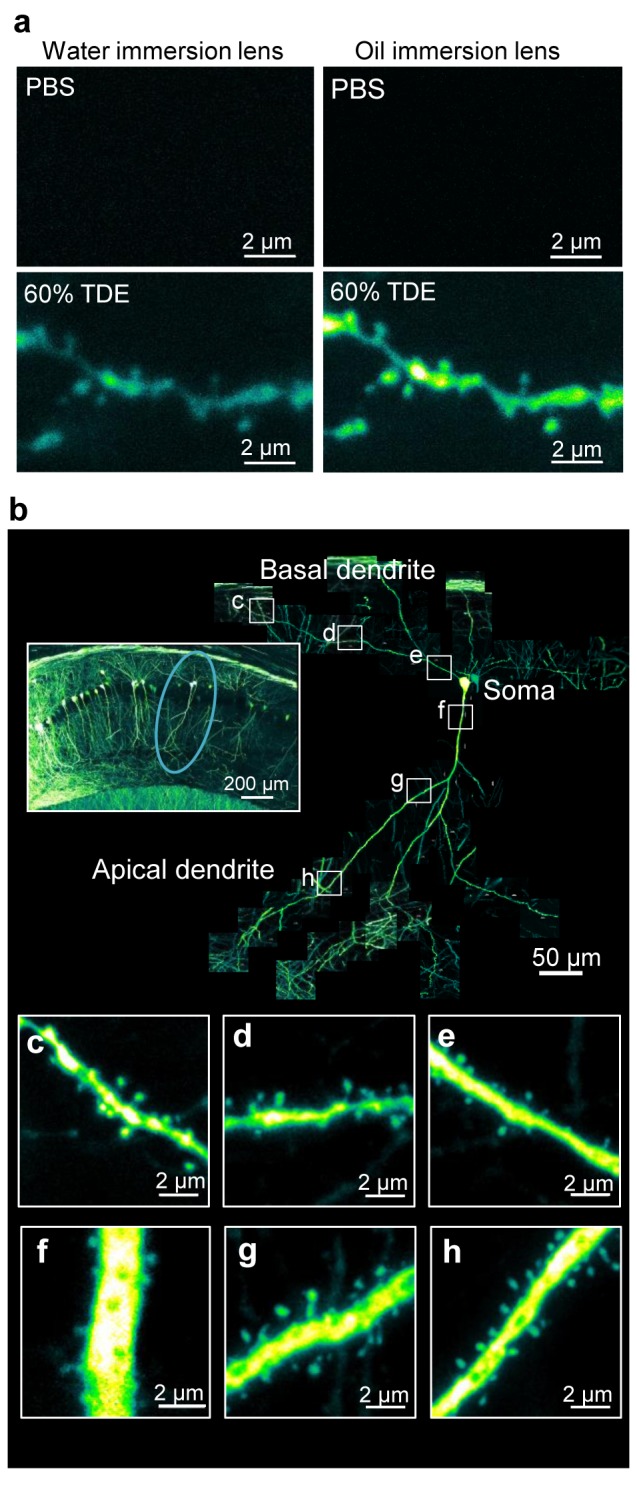
(a) Combination of TDE treatment and water/oil-immersion objective lens with a high NA for imaging dendritic spine shapes in deep (100 µm) regions in a fixed brain slice. (b) Connected images of dendritic spines along single pyramidal neurons extending from a depth of 30 to 100 μm from the surface of the hippocampal slice. The left inset shows a low-magnification image of the hippocampus, and the circle shows the observed neuron. (c-h) Magnified images of the dendritic spine shapes on the basal dendrite (c-e) and the apical dendrite (f-h) along the neuron shown in (b). All images are maximum projection images.