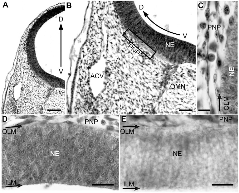
Figure 3. Radial organization of the OT vasculature during DS1 (ED3; HH20).
(D-V sections; a-d: H-E; e: Diaphorase). (A-B) The OT is surrounded by the perineural vascular plexus (PNP). (C) Higher magnification of the box shown in b. Some endothelial cells contact the outer limiting membrane. (D-E) Endothelial cells are closely attached to the neuroepithelium basal surface but the OT wall is still deprived of vessels (Bars: A: 100 µm; B: 50 µm; C: 10 µm; D-E: 25 µm).

