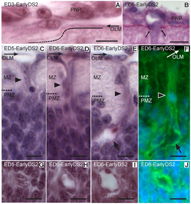
Figure 5. Ingression of primitive vessels into the OT (Early DS2; ED3–6; HH20–29).
(A-D and F-H: H-E; E and I: Notch and Hoechst). (A) Endothelial cells attach to the outer limiting membrane (OLM) and degrade the basal membrane (dotted line). (B) PAS-Stained section reveals the interruption (between arrows) of the basement membrane at a site where a vessel sprouts traverses the pial surface and penetrates the neuroepithelium. (C) Solid sprouts (arrowhead) from the perineural vascular plexus (PNP) penetrate the neuroepithelium and pass through the marginal zone (MZ) toward the premigratory zone (PMZ). (D) Early primitive solid radial vessel (arrowhead) entering the PMZ. (E-F) After entering the PMZ the tip of the growing vessel develops a lumen (arrows). (G-J) Transverse sections of primitive radial vessels in different stages of development (Bars: A: 5 µm; B-J: 10 µm).

