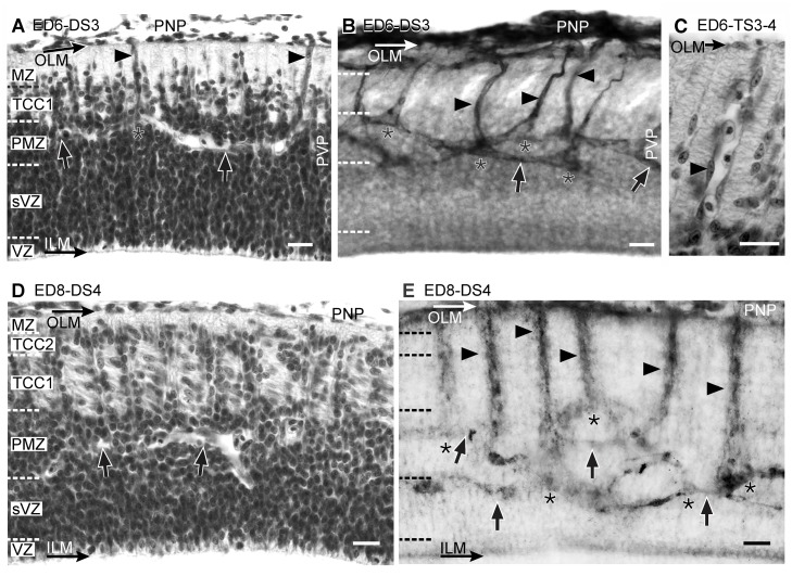
Figure 7. Radial organization of the OT vasculature during DS3 (A-B, ED6; HH29), TS3–4 (C, ED6, HH29) and DS4 (D-E: ED8; HH34).
(A, C and D: H-E; B and E: Diaphorase). (A) The TCC1 appears between the marginal zone (MZ) and the premigratory zone (PMZ). (B) The primitive radial vessels (arrowheads) traverse the TCC1 and form the periventricular plexus (PVP). The tangential vessels of the PVP (arrows) display two preferential positions: most of them run through the TCC1-PMZ interface or through the PMZ-SVZ interface. Some of them run from one interface to the other. Asterisks: points of bifurcation. (C) During the TS-3–4, the primitive radial vessels (arrowhead) increase in length and diameter and are frequently occupied by several red blood cells (D) The TCC2 appears between the marginal zone (MZ) and the TCC1. Columns of radially migrating neurons can be seen at regular intervals traversing the TCC1. TCC1 neurons can be identified by their tangential orientation. (E) The OT cortex thickening is accompanied by the elongation of the radial vessels (arrowheads). Arrows: tangential vessels of the periventricular plexus; asterisks: bifurcations (Bars: 20 µm).

